Common Remake Solutions
Follow the tips listed below to help prevent remakes.
-
1Tight Contacts
- Check for a bulge below the ideal contact area on the crown. Smooth down the bulge if necessary.
- Check contact areas on the adjacent teeth, looking for draw or bulges that can be smoothed out.
-
2Light ContactsIf the contact is light/open, verify margins, fit, and shade
Evaluate adjacent contact areas and adjust as necessary:- Parallel with the prep and with each other for draw
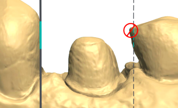
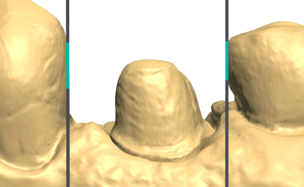
- Broad contacts for ideal restoration(s)
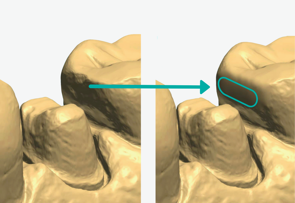
- Rescan the prep and adjacent teeth only. (Note: tissue retraction is not necessary if the crown margins and fit were good.)
See step 4 of our Normal Case Protocol to see more details on which tools we recommend for contact adjustment. -
3Short or Open Margins
- Double pack cord before scanning.
- If necessary, use a laser to expose the margins.
- Make sure the area is completely clean and dry, then rescan.
- For the best scanning results, leave both cords in for 5 minutes then remove the top cord (#02) immediately before scanning. Leave the bottom cord (#00) in while scanning.

-
4Incorrect Shade
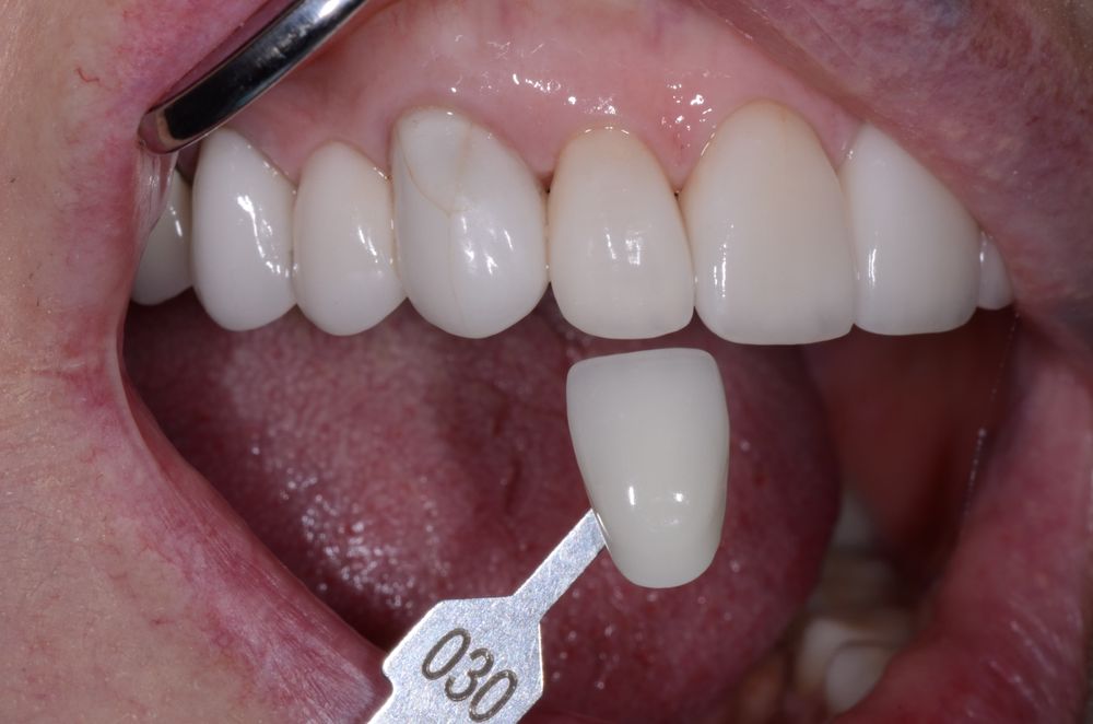
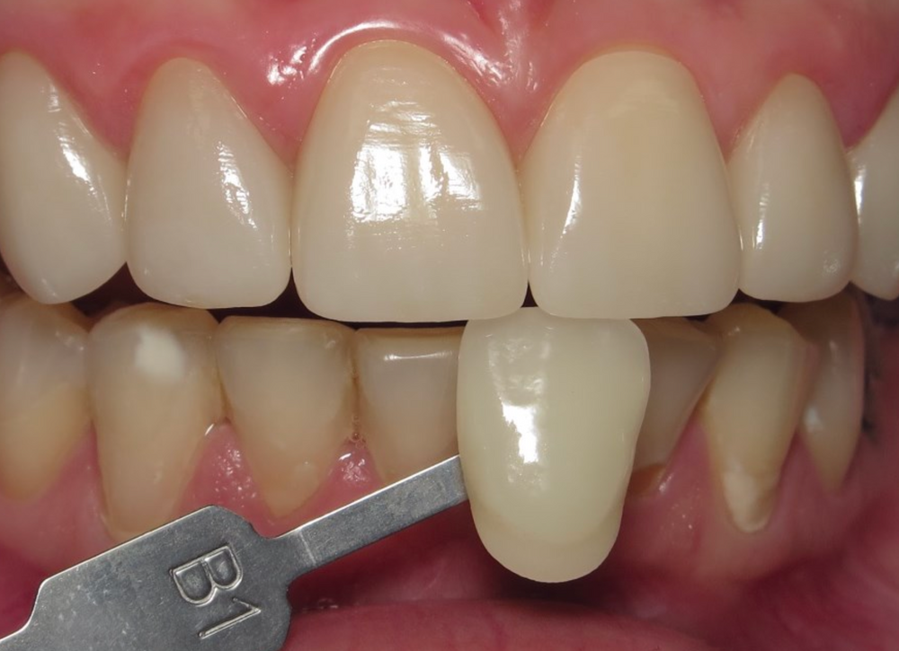
- Send photos of the adjacent teeth with a full shade tab in view. The incisal edge of the shade tab should be lined up with the incisal edge of a tooth for the most accurate results. Watch out for bad glare.
- Provide additional information (calcification, incisal translucency, etc.) to help us accurately match the shade.
-
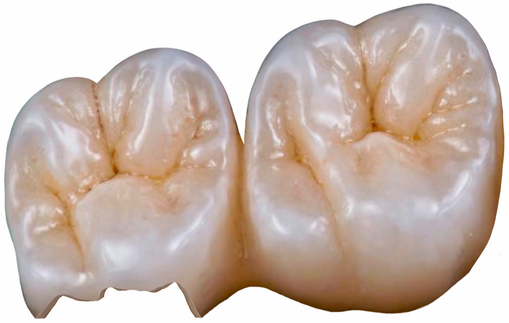
5FracturesTo assist us in determining a potential cause and solution for the fracture, please:
- inspect the prep for sharp edges.
- inspect the bite for sufficient clearance (we suggest 1.5mm).
- rescan the prep and bite and send us the new scans.
-
6Esthetic Issues
- Provide precise information regarding design preferences.
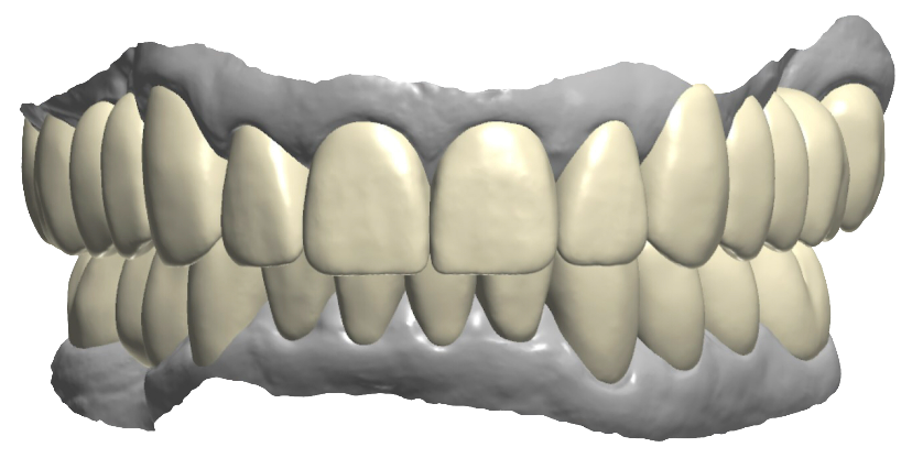
- Send photos and any additional notes with the case. Consider including drawings of your desired results (see example below).
- For highly esthetic cases, request screenshots or a 3D preview before the crowns are milled.

-
7Open Occlusion
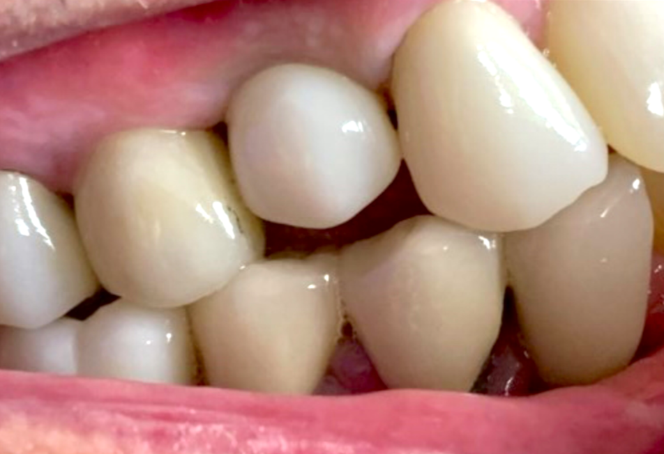
- Ensure the patient is biting in centric occlusion.
- Take a new bite scan and evaluate the alignment by comparing the scan to the patient's mouth.
- For implant cases:
- take an x-ray to confirm the scan body is fully seated.
- remove the scan body before taking the bite scan.
Did this answer your question?
If you still have a question, we’re here to help. Contact us


